Groupleader: Sebastian Schuck

Sebastian Schuck
Organelle Homeostasis

Research
Organelle Homeostasis
Biogenesis and Degradation of the Endoplasmic Reticulum
We want to elucidate how cells ensure homeostasis of the endoplasmic reticulum (ER). We are asking questions such as: How do cells adjust ER size to physiological demand? How do cells control ER shape to optimally support its functions? How do cells eliminate ER damage? Using budding yeast and human cells, we focus on ER membrane biogenesis, which enables organelle expansion and remodelling and helps to prevent an accumulation of misfolded proteins. Two other processes we have investigated are ER-phagy, which mediates autophagic organelle degradation, and SHRED, which regulates proteasomal degradation of misfolded cytosolic and ER membrane proteins.
The ER is a morphologically complex organelle with vital functions in protein folding and lipid synthesis. When the ER is unable to fold its load of newly synthesized polypeptides, misfolded proteins accumulate and cause ER stress. Misfolded proteins activate the unfolded protein response (UPR), which increases the protein folding capacity of the ER and triggers massive expansion of the ER membrane, both in yeast (Figure 1) and in human cells (Figure 2). We have identified genes required for ER membrane expansion and determined how they regulate lipid metabolism (Papagiannidis, Bircham et al, 2021). Furthermore, we have found that protein relocalization is a major aspect of cellular reorganization in response to ER stress (Platzek et al, 2025).
Secretory cells, such as antibody-secreting plasma cells, need to expand the ER membrane during differentiation. Therefore, finding out how cells adjust ER size will help us understand how cells respond to stress and also how they differentiate. Besides the regulation of ER size, we are keen to understand how cells control ER shape, including the proportion of ER tubules and sheets, the generation of ER stacks and the formation of ER whorls.

Figure 1. ER membrane expansion in yeast. Cells expressing Sec63-GFP to highlight the cytoplasmic ER (cER) and the nuclear envelope (NE). Cells exposed to ER stress have a vastly expanded cytoplasmic ER.

Figure 2. ER membrane expansion in human cells. Tissue culture cells expressing RFP-KDEL to highlight the ER. Cells exposed to ER stress convert their tubular ER network into sheet-like ER.
ER-phagy
Autophagy (cellular self-eating) is another response to ER stress. Upon stress, cells turn on selective autophagy of the ER, which can occur by macroautophagy and microautophagy. We have explored micro-ER-phagy, which in yeast involves a spectacular ER restructuring that gives rise to multilamellar whorls. These whorls are then sent to the lysosome for degradation (Figure 3). We have shown that micro-ER-phagy does not require the well-known core autophagy machinery but depends on ESCRT proteins (Schäfer et al., 2020). Through micro-ER-phagy, cells may sacrifice parts of their ER to destroy protein aggregates. Moreover, when stress has been resolved, micro-ER-phagy can downsize the ER and reverse organelle expansion. In this way, the UPR and ER-phagy work together to refold or degrade damaged proteins, and to expand or shrink the ER as needed. Hence, ER-phagy helps to maintain ER homeostasis and may be relevant for diseases related to ER function, such as cancer and diabetes.

Figure 3. Correlative light and electron microscopy of micro-ER-phagy in yeast. Micro-ER-phagy can be triggered by expression of an artificial transmembrane protein called 'ER-phagy inducer'. Fluorescence images show a ring-shaped structure positive for a general ER marker and the ER-phagy inducer. The corresponding electron micrograph reveals that this structure is a large multilamellar ER whorl inside the yeast lysosome.
SHRED
Protein folding is error-prone, especially during stress. Cells possess elaborate quality control machinery, including numerous chaperones and ubiquitin ligases, to promote proper folding and degrade folding failures. Stress responses like the UPR tune quality control to current demand. We have uncovered a novel stress response pathway termed SHRED, for stress-induced homeostatically regulated protein degradation (Figure 4; Szoradi et al, 2018; Peters, Kanngießer et al, 2025). SHRED is activated when stress stimulates transcription of the Roq1 gene. The Roq1 protein is cleaved by the protease Ynm3. Truncated Roq1 then binds to the ubiquitin ligase Ubr1 as a pseudosubstrate, reprograms Ubr1's substrate specificity and directs it towards misfolded cytosolic and ER membrane proteins. The resulting more stringent quality control enhances stress resistance. Deteriorating protein quality control during aging is a key factor for the onset of neurodegenerative diseases such as Alzheimer’s. Moreover, cancer cells suffer from chronic folding stress and depend on heightened quality control for survival.

Figure 4. SHRED. Under non-stress conditions, the ubiquitin ligase Ubr1 degrades proteins with positively charged N-terminal residues as part of the N-degron pathway (left). Under stress conditions, Roq1 is produced, is cleaved by Ynm3 and binds to Ubr1 as a pseudosubstrate. This reprograms Ubr1 and stimulates the degradation of misfolded proteins (right).
Selected publications
Platzek A, Odehnalova K, Schessner JP, Borner GH, Schuck S (2025) Dynamic Organellar Mapping in yeast reveals extensive protein localization changes during ER stress. Nature Communications (abstract)
Peters N*, Kanngießer S*, Pajonk O, Salazar Claros R, Hubbe P, Mogk A, Schuck S (2025) Reprograming of the ubiquitin ligase Ubr1 by intrinsically disordered Roq1 through cooperating multifunctional motifs. EMBO Journal (abstract)
Papagiannidis D*, Bircham PW*, Lüchterborg C, Pajonk O, Ruffini G, Brügger B, Schuck S (2021) Ice2 promotes ER membrane biogenesis in yeast by inhibiting the conserved lipin phosphatase complex. EMBO Journal (abstract)
Schäfer JA, Schessner JP, Bircham PW, Tsuji T, Funaya C, Pajonk O, Schaeff K, Ruffini G, Papagiannidis D, Knop M, Fujimoto T, Schuck S (2020) ESCRT machinery mediates selective microautophagy of endoplasmic reticulum in yeast. EMBO Journal (abstract)
Szoradi T, Schaeff K, Garcia-Rivera EM, Itzhak DN, Schmidt RM, Bircham PW, Leiss K, Diaz-Miyar J, Chen VK, Muzzey D, Borner GH, Schuck S (2018) SHRED is a regulatory cascade that reprograms Ubr1 substrate specificity for enhanced protein quality control during stress. Molecular Cell (abstract)
CV
since 2021 Professor for Biochemistry and Molecular Cell Biology
Heidelberg University Biochemistry Center
2013-2021 Independent group leader
Center for Molecular Biology at Heidelberg University
2006-2013 Postdoctoral fellow with Peter Walter
University of California, San Francisco
2001-2006 PhD student and postdoctoral fellow with Kai Simons
Max Planck Institute of Molecular Cell Biology, Dresden
1995-2000 Biochemistry student
Universities of Hannover and Tübingen
Publications
bioRxiv (abstract)
Pajonk O*, Albert L*, Schäfer JA, de Jager L, Martin de Hijas C, Papagiannidis D, Odehnalova K, Friemel N, Esch BM, Fröhlich F, Luzarowski M, Borner GHH, Förster F, Schuck S (2026)
Systematic evaluation of tools for auxin-inducible protein degradation in budding yeast.
Molecular Biology of the Cell (abstract)
Hubbe P, Sharma C, Pajonk O, Peters N, Guschtschin-Schmidt N, Friemel N, Schuck S (2026)
Uncovering the initial response: Intra-mitochondrial surveillance activates the UPRmt.
Molecular Cell (abstract)
Taskin AA, Shankar S, Peselj C, Flotho A, Gomez-Fabra Gala M, Poveda-Huertes D, Myketin L, Mutlu D, Marada A, Schuck S, Jeske M, Büttner S, Luzarowski M, Meisinger C, Vögtle FN (2026)
Dynamic Organellar Mapping in yeast reveals extensive protein localization changes during ER stress.
Nature Communications (abstract)
Platzek A, Odehnalova K, Schessner JP, Borner GH, Schuck S (2025)
Reprograming of the ubiquitin ligase Ubr1 by intrinsically disordered Roq1 through cooperating multifunctional motifs.
EMBO Journal (abstract)
Peters N*, Kanngießer S*, Pajonk O, Salazar Claros R, Hubbe P, Mogk A, Schuck S (2025)
Ice2 promotes ER membrane biogenesis in yeast by inhibiting the conserved lipin phosphatase complex.
EMBO Journal (abstract)
Papagiannidis D*, Bircham PW*, Lüchtenborg C, Pajonk O, Ruffini G, Brügger B, Schuck S (2021)
ESCRT machinery mediates selective microautophagy of endoplasmic reticulum in yeast.
EMBO Journal (abstract)
Schäfer JA, Schessner JP, Bircham PW, Tsuji T, Funaya C, Pajonk O, Schaeff K, Ruffini G, Papagiannidis D, Knop M, Fujimoto T, Schuck S (2020)
ESCRTing endoplasmic reticulum to microautophagic degradation (commentary).
Autophagy (abstract)
Schäfer JA, Schuck S (2020)
Microautophagy - distinct molecular mechanisms handle cargoes of many sizes (review).
Journal of Cell Science (abstract)
Schuck S (2020)
The proteasome biogenesis regulator Rpn4 cooperates with the unfolded protein response to promote ER stress resistance.
eLife (abstract)
Schmidt RM, Schessner JP, Borner GH, Schuck S (2019)
SHRED is a regulatory cascade that reprograms Ubr1 substrate specificity for enhanced protein quality control during stress.
Molecular Cell (abstract)
Szoradi T, Schaeff K, Garcia-Rivera EM, Itzhak DN, Schmidt RM, Bircham PW, Leiss K, Diaz-Miyar J, Chen VK, Muzzey D, Borner GH, Schuck S (2018)
Biogenese und Autophagie des Endoplasmatischen Retikulums (review).
BIOspektrum (abstract)
Schäfer JA, Schuck S (2017)
Guidelines for the use and interpretation of assays for monitoring autophagy (review).
Autophagy (abstract)
Klionsky DJ, Abdelmohsen K, ..., Schuck S, ... Zughaier SM (2016)
On keeping the right ER size (commentary).
Nature Cell Biology (abstract)
Schuck S (2016)
ER-phagy mediates selective degradation of endoplasmic reticulum independently of the core autophagy machinery.
Journal of Cell Science (abstract)
Schuck S, Gallagher CM, Walter P (2014)
Quantitative analysis and modeling of katanin function in flagellar length control.
Molecular Biology of the Cell (abstract)
Kannegaard E, Rego EH, Schuck S, Feldman JL, Marshall WF (2014)
Seg1 controls eisosome assembly and shape.
Journal of Cell Biology (abstract)
Moreira KE*, Schuck S*, Schrul B, Fröhlich F, Moseley JB, Walther TC, Walter P (2012)
BAX inhibitor-1 regulates autophagy by controlling the IRE1α branch of the unfolded protein response.
EMBO Journal (abstract)
Castillo K, Rojas-Rivera D, Lisbona F, Caballero B, Nassif M, Court FA, Schuck S, Ibar C, Walter P, Sierralta, J, Glavic A, Hetz C (2011)
Homeostatic adaptation to endoplasmic reticulum stress depends on Ire1 kinase activity.
Journal of Cell Biology (abstract)
Rubio C, Pincus D, Korennykh A, Schuck S, El-Samad H, Walter P (2011)
Role of EBAG9 protein in coat protein complex I-dependent glycoprotein maturation and secretion processes in tumor cells.
FASEB Journal (abstract)
Wolf J, Reimer T, Schuck S, Rüder C, Gerlach K, Müller EC, Otto A, Dörken B, Rehm A (2010)
Generation of cubic membranes by controlled homotypic interaction of membrane proteins in the endoplasmic reticulum.
Journal of Biological Chemistry (abstract)
Lingwood D, Schuck S, Ferguson C, Gerl MJ, Simons K (2009)
Membrane expansion alleviates endoplasmic reticulum stress independently of the unfolded protein response.
Journal of Cell Biology (abstract)
Schuck S, Prinz WA, Thorn KS, Voss C, Walter P (2009)
Morphological homeostasis by autophagy (commentary).
Autophagy (abstract)
Lingwood D, Schuck S, Ferguson C, Gerl MJ, Simons K (2009)
Depletion of apical transport proteins perturbs epithelial cyst formation and ciliogenesis.
Journal of Cell Science (abstract)
Torkko JM, Manninen A, Schuck S, Simons K (2008)
ER-phagy: selective autophagy of the endoplasmic reticulum (commentary).
Autophagy (abstract)
Bernales S*, Schuck S*, Walter P (2007)
Rab10 is involved in basolateral transport in polarized MDCK cells.
Traffic (abstract)
Schuck S*, Gerl MJ*, Ang AL, Manninen A, Keller P, Mellman I, Simons K (2007)
Controversy fuels trafficking of GPI-anchored proteins (commentary).
Journal of Cell Biology (abstract)
Schuck S, Simons K (2006)
Detergent-resistant membranes and the use of cholesterol depletion. In Cell Biology: A Laboratory Handbook, Volume 2. J. E. Celis, editor. Elsevier Science, USA. Book chapter (full text)
Schuck, S., Honsho, M., & Simons, K. (2006)
Generation of single and double knockdowns in polarized epithelial cells by retrovirus-mediated RNA interference.
Proceedings of the National Academy of Sciences USA (abstract)
Schuck S*, Manninen A*, Honsho M, Füllekrug J, Simons K (2004)
Polarized sorting in epithelial cells: raft clustering and the biogenesis of the apical membrane (review).
Journal of Cell Science (abstract)
Schuck S, Simons K (2004)
Resistance of cell membranes to different detergents.
Proceedings of the National Academy of Sciences USA (abstract)
Schuck S*, Honsho M*, Ekroos K, Shevchenko A, Simons K (2003)
The kinase MSK1 is required for induction of c-fos by lysophosphatidic acid in mouse embryonic stem cells.
BMC Molecular Biology (abstract)
Schuck S, Soloaga A, Schratt G, Arthur JS, Nordheim A (2003)
Serum response factor is required for immediate-early gene activation yet is dispensable for proliferation of embryonic stem cells.
Molecular and Cellular Biology (abstract)
Schratt G, Weinhold B, Lundberg A, Schuck S, Berger J, Schwarz H, Weinberg R, Ruther U, Nordheim A (2001)
Current Lab Members
| Sebastian Schuck 2013- |
 |
Sebastian still fantasises about doing experiments himself, but the harsh reality is that he is stuck at the desk. |
| Saccharo Cerevis 2013- |
 |
Saccharo has been our resident oracle since the lab started in July 2013. What s/he says is mostly incomprehensible but sometimes reveals amazing insights. |
| Oliver Pajonk 2018- |
 |
Oli did his Master's and PhD with us and is now a postdoc to investigate ESCRTs in ER remodelling. He enjoys music, running and convincing other lab members to join him for running events. |
| Anke Sander 2021- |
 |
Anke is our administrative assistant. She fights her way through the bureaucratic jungle, always building bridges between science and administration – and also between duty, coffee and cake. |
| Rolf Schmidt 2022- |
 |
Rolf left us after completing his PhD in 2019 but then thought better of it and returned as postdoc. His guitar collection has been growing all the while. |
| Petra Hubbe 2022- |
 |
Petra joined us as a technician after many previous jobs, including at the ZMBH. She keeps our many toys for Saccharo in good shape, teaches Saccharo new tricks, and finds out more about what s/he does. |
| Natalie Friemel 2023- |
 |
Natalie joined the lab in June 2023 for her Master thesis and then stayed as a PhD student. When not investigating ER morphology in yeast, she enjoys hiking, running and her arts & crafts projects. |
| Klára Odehnalová 2023- |
 |
Klára started her PhD in Octocber 2023. Having quickly made friends with Saccharo, she wants to find out how the ER changes its shape. Outside the lab, her many interests include strange critters, such as bearded dragons. |
| Sneha Bhatt 2024- |
 |
Sneha joined the lab as a PhD student in January 2024 and is determined to find out how ER morphogenesis works in human cells. Sneha self-identifies as a science geek but also enjoys travelling and cooking (although the right spices are currently a bit hard to come by). |
| Heike Adler 2025- |
 |
Heike joined us as a technician and in this way returned home to the BZH. She helps us get better at studying ER morphogenesis in mammalian cells. |
| Pauline Terrien 2025- |
 |
Pauline arrived in Heidelberg in November 2025 to find out how ER size is controlled in mammalian cells. |
| Leanne de Jager 2026- |
 |
Leanne did her PhD with Friedrich Förster in Utrecht, where she became an expert in cryo-EM. She still loves electron microscopy but now broadened her approach to study ER dynamics in mammalian cells. Leanne also is an avid runner. |
| Aditi Yadav 2026- |
 |
Aditi is doing her Master's thesis with us. She wants to find out if starvation-induced autophagy in yeast really is non-selective or if some proteins are chewed up preferentially. |
Former Lab Members
| Tamas Szoradi 2013-2018 |
 |
In his PhD, Tamas unravelled SHRED in all its glory. He was also the first of many boldering experts in the lab. Tamas then moved to New York to do a postdoc with Liam Holt. |
| Jasmin Schäfer 2014-2020 |
 |
Jasmin's PhD was all about whorls. The force was strong with this one and Jasmin heroically cracked (some of) the secrets of micro-ER-phagy. She then moved to Frankfurt to do a postdoc with Christian Münch. |
| Dorottya Polos 2014-2015 |
 |
Dorottya was an ERASMUS student with us. Afterwards, she returned to London to complete her Bachelor’s degree and do her PhD with Margaret Dallman. |
| Rolf Schmidt 2014-2019 |
 |
In his PhD, Rolf investigated how yeast cells adapt protein degradation capacity to stress. He then stayed in Heidelberg to do a postdoc in Neuroscience with Carlos Bas Orth. |
| Peter Bircham 2015-2018 |
 |
Peter was a postdoc with us and studied ER size control. Outside the lab, Peter still put yeast to good use for brewing. This naturally led to his second postdoc with Kevin Verstrepen in Leuven, where Peter engineered yeast for making better beer. |
| Verena Bittl 2015 |
 |
Verena did her Master thesis with us to explore potential ER-phagy substrates. She then moved to Frankfurt to pursue her PhD with Anja Bremm. |
| Dimitris Papagiannidis 2016-2022 |
 |
Dimitris did his Master thesis with us and then his PhD. By climbing from tubules to sheets and doing extensive bouldering on the yeast ER, he figured out what Ice2 actually does. He then ventured up another local hill to do a postdoc with Nassos Typas and Mikhail Savitski at EMBL. |
| Julia Schessner 2017-2018 |
 |
Julia did her Master thesis with us on visualizing and quantifying micro-ER-phagy by time lapse microscopy. For her PhD, Julia joined Georg Borner’s group at the MPI of Biochemistry in Munich. |
| Giulia Ruffini 2018-2025 |
 |
Giulia did her Master thesis and her PhD with us to study ER biogenesis in human cells. Her efforts brought the beauty of the mammalian ER to the lab, and she was the first to capture a picture of the fabled science fairy. Giulia then took a lab manager position at EMBL. |
| Sibylle Kanngießer 2019-2024 |
 |
Sibylle's PhD was a close collaboration with Niklas to unravel exactly how Roq1 talks to Ubr1 to activate SHRED. She then moved to Hamburg to take an industry position. |
| Niklas Peters 2019-2025 |
 |
Niklas worked closely with Sibylle on the mechanism of SHRED. He stayed on as a postdoc for a little bit before taking an industry job in nearby Bensheim. When Niklas left the Äkta wept. |
| Lis Albert 2020-2025 |
 |
Lis spent her Master thesis and her PhD to find out how ESCRT proteins talk with the ER. She closely collaborated with Oli, both on science and also on many other projects such as running and bouldering. She then moved on to a science communication traineeship at EMBL, joining Giulia and Dimitris. |
| Ayelén Valko 2020-2022 |
 |
Ayelén was a postdoc with us, working on micro-ER-phagy and ER morphogenesis. Aye is a serious artist with a (e)special interest in science-inspired paintings. She decided to turn to art full time and you can visit her work at www.ayelenvalko.com. |
| Carlos Martin de Hijas 2021 |
 |
Carlos did his Master thesis to look at transcriptional responses to ER stress, mostly by staring at the computer screen. Carlos then took a job at a local Biotech company. |
| Anna Platzek 2021-2025 |
 |
For her PhD, Anna initially wanted to find out what ER whorls are made of and ended up discovering how the entire yeast proteome is remodelled during stress. |
| Inge Reckmann 2021-2024 |
 |
Inge was a long-time technician of Felix Wieland before we moved into the lab space she had been keeping in good shape for many years. After three happy years with us, we are sad to lose her to retirement. |
| Rafael Salazar Claros 2022 |
 |
Rafael did his Master thesis by joining the SHREDders. He then continued studying the ubiquitin proteasome system as a PhD student in Satpal Virdee's lab in Dundee. |
| Alexander Wirth 2022-2023 |
 |
Alex did his Master thesis with us to figure out how reticulon proteins control ER morphology in yeast. He then joined Jirka Peschek's group at our institute for his PhD. |
| Adam Jamalov 2026 |
 |
Adam studies Bioinformatics and Computational Biology at Université Côte d’Azur and joined us as an ERAMUS student. Through some machine learning magic and the creation of the YeastBuddy app, he help us get better at quantitative image analysis. |
Lab Pictures

February 2026. The lab on a fabulous trip to an exotic ... local greenhouse. From left to right: Oli, Natalie,
Petra (with Saccharo), Leanne, Sneha (with a tissue culture cell featuring a pink ER), Adam, Pauline,
Sebastian, Klara and Heike.
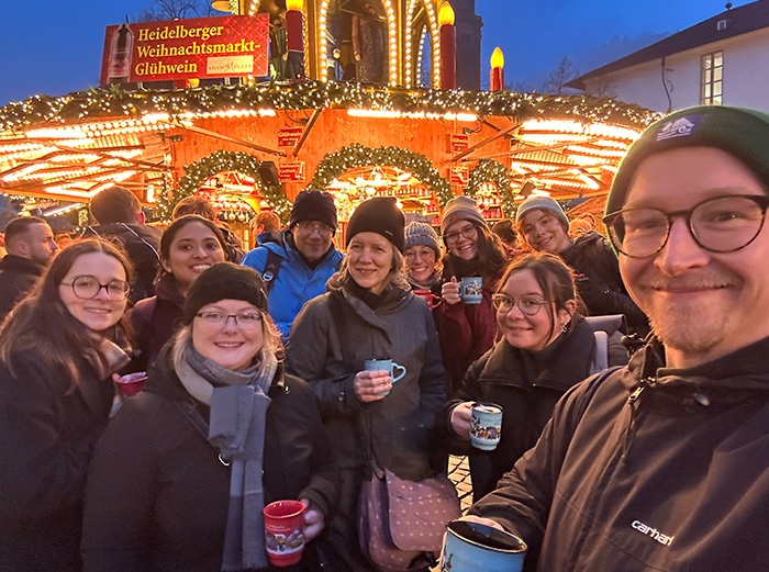
December 2025. Our annual trip to the Heidelberg Christmas market, of course involving Glühwein.
From left to right: Pauline, Sneha, Anke, Sebastian, Heike, Anna, Lis, Klara, Nathalie, Oli.
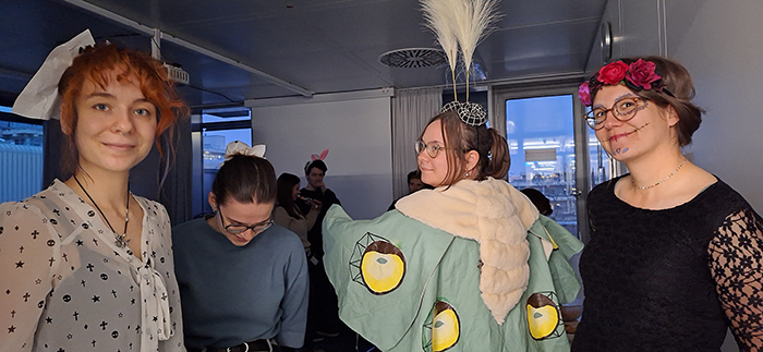
November 2025. At the Halloween party during Beer & Science. From left to right: Natalie, Pauline, Klara
(as a moth) and Lis.

October 2025. Team building at the lab retreat. Being able to solve problems without knowing what you
are doing sure is a helpful skill for research. From left to right: Pauline, Sneha, Sebastian, Leanne, Natalie,
a tiny piece of Oli, Lis and Heike. And a cat wondering what those humans are doing.

December 2024. Three graduations in one months: Niklas, Sibylle and Oli all successfully defended their
PhDs - congratulations!

October 2024. Oli grinding coffee at an amazing speed. And thus preventing our lab retreat from grinding
to an undercaffeinated halt.

September 2024. Look, someone did a graffiti of a cell! From left to right: Sneha, Natalie, Klara, Sebastian
(cradling Saccharo, who is busy budding), Lis, Petra, Oli, Sibylle (who appears to be pointing at the wrong
organelle), Niklas, Giulia and Anna.

October 2023. Drawing octopuses at the lab retreat. Hard at work from left to right: Natalie (whose hair
is on fire), Petra, Sebastian, Klara, Niklas and Inge.

January 2023. The lab in its new home. From left to right: Anke, Rolf, Sibylle, Lis, Alex, Petra, Oli, Inge, Anna, Sebastian, Giulia, Saccharo and Niklas.

December 2022. Preparing a talk can be tough. And confusing. Just ask Rolf.

May 2022. Dimitris graduates! In keeping with local traditions, a silly hat is made (the proud hatters are
Niklas, Rolf, Lis and Anna) ...

... and then the graduate is made to look like a muppet (Dimitris
surrounded by fellow Schuck lab graduates, Jasmin, Rolf and Tamas).

March 2022. From left to right, back row: Ayelèn, Inge, Sibylle, Dimitris; middle row: Niklas, Sebastian, Lis,
Giulia, Natalie; front row: Anke, Anna, Oli.

August 2020. The lab in its natural environment.
From left to right: Oli, Niklas, Dimitris, Sebastian, Giulia, Sibylle, Lis and Uxia.

March 2020. Lab meeting in times of Corona.

February 2020. Jasmin has defended her PhD and even the dark side pays its respects.

September 2019, in deep thought at the lab retreat. From left to right: Jasmin, Giulia, Dimitris, Sebastian,
Niklas (apparently having an idea), Sibylle and Oli.

July 2019, the lab participates in the 10 K run of the National Center for Tumor Diseases. From left to
right: Oli, Niklas, Dimitris, Sebastian and Sibylle (we blame the red faces on the camera).

October 2018. From left to right: Oli, Sebastian, Giulia, Dimitris, Rolf, Jasmin and Carlos.
Open Positions
In addition, we are currently looking for a new administrative assistant, please see here:
Contact
Biochemistry Center (BZH)
Im Neuenheimer Feld 328
69120 Heidelberg
